As an experienced ophthalmologist in Lagos with over five years of clinical practice, I provide an expert, detailed analysis of hypertensive retinopathy, its symptoms, stages, treatment options, and practical steps for effective management.
Understanding the Growing Threat of Hypertensive Retinopathy
Hypertension has become a silent epidemic across Nigeria, particularly in urban centres like Lagos, where stress, dietary changes, and lifestyle factors have contributed to its increasing prevalence. As a clinician who has managed hundreds of hypertensive patients over the past five years, I’ve observed a disturbing trend: a growing number of these individuals also develop complications affecting their vision — one of the most serious being hypertensive retinopathy.
Hypertensive retinopathy is a condition in which elevated blood pressure damages the tiny blood vessels of the retina, the light-sensitive tissue at the back of the eye. Left unchecked, this condition can lead to irreversible vision loss, significantly impairing quality of life.
In my clinic, patients often present late, when symptoms are already advanced. This delay often results from unawareness. Many Lagos residents understand hypertension as a “blood pressure issue,” yet few realise that it can quietly steal their sight.
The problem, therefore, is twofold:
- Lack of awareness about the ocular effects of hypertension.
- Delayed diagnosis and treatment of hypertensive retinopathy.
Hypertensive retinopathy is not only a medical concern but a public health issue — one that requires both patient education and expert intervention.
How Hypertension Damages the Retina
To address this problem effectively, it is essential to understand the mechanism and progression of hypertensive retinopathy.
When blood pressure remains consistently elevated, it exerts excessive force on the delicate walls of retinal blood vessels. Over time, this causes narrowing, thickening, and leakage in these vessels. The retina, deprived of adequate oxygen and nutrients, begins to deteriorate.
The Four Stages of Hypertensive Retinopathy
A clear understanding of disease staging is crucial both for diagnosis and management. Based on the Keith-Wagener-Barker classification, the four stages of hypertensive retinopathy are:
- Stage 1 – Mild Retinal Arteriolar Narrowing:
The earliest stage involves subtle narrowing of the retinal arteries. The patient may not notice any symptoms at this point, but the ophthalmologist can detect these changes during a fundus examination. - Stage 2 – More Pronounced Narrowing and Arteriovenous (AV) Nicking:
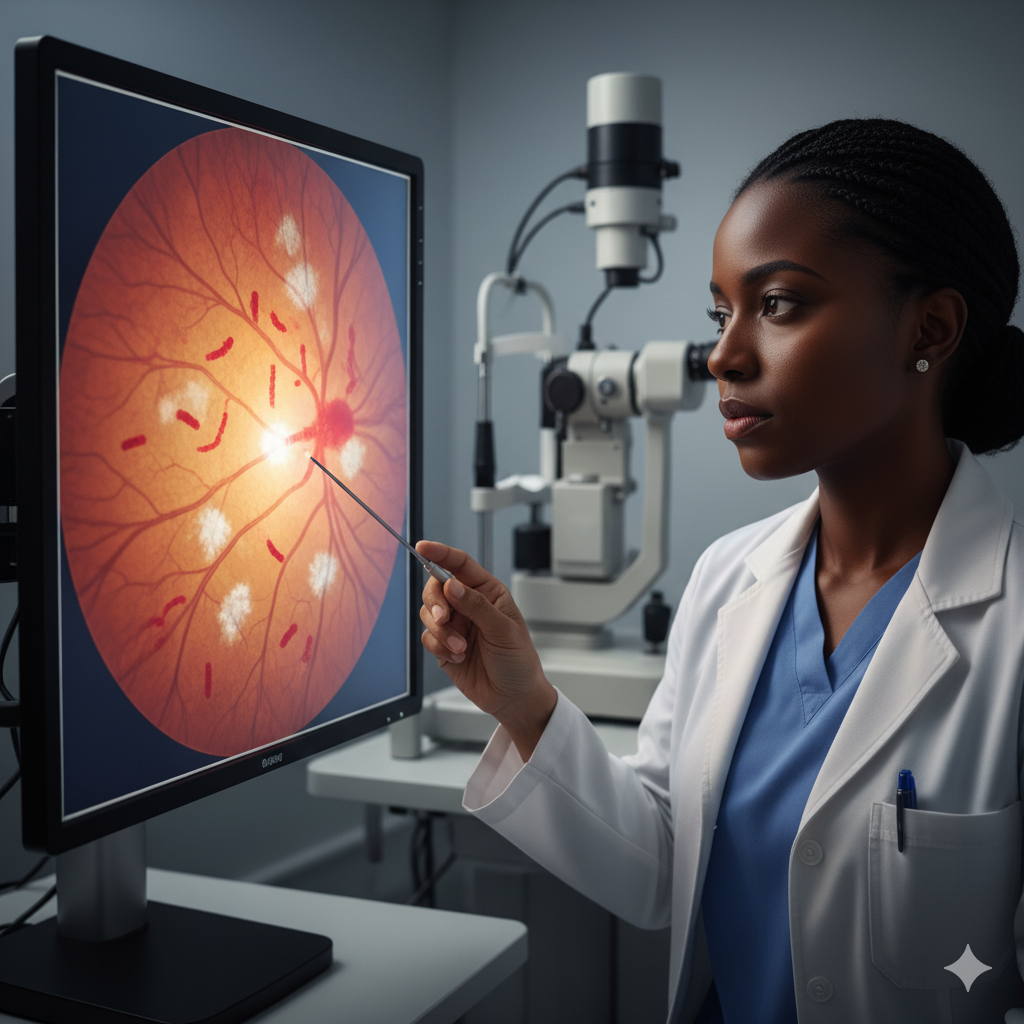
As hypertension persists, the vessel walls thicken, compressing nearby veins — a finding known as AV nicking. This indicates sustained pressure damage. - Stage 3 – Retinal Hemorrhages and Exudates:
The damaged vessels begin to leak blood and fluid. On examination, flame-shaped hemorrhages, cotton wool spots, and hard exudates may appear. At this stage, vision disturbances often begin. - Stage 4 – Malignant (Severe) Hypertensive Retinopathy:
This is the most dangerous phase, characterised by optic disc swelling (papilledema). It signifies a medical emergency requiring immediate intervention to prevent permanent vision loss or even stroke.
The Four Signs of Hypertensive Retinopathy
Clinically, the four signs of hypertensive retinopathy that patients and clinicians should be alert to include:
- Blurred Vision – Caused by retinal swelling or haemorrhage.
- Headaches – Resulting from increased intracranial or ocular pressure.
- Double Vision or Visual Distortion – Due to irregularities in retinal structure.
- Gradual Vision Loss – Often indicating advanced vascular damage.
It’s worth noting that these symptoms may develop gradually. In many Lagos patients I’ve managed, the first noticeable complaint is a vague “fog” or blurriness, a symptom often ignored until significant vision impairment occurs.
Risk Factors and Local Context
In Lagos, common contributing factors include:
- Poor hypertension control due to inconsistent medication use.
- Sedentary lifestyles and diets high in salt and processed foods.
- Limited routine eye examinations, especially among adults above 40.
Understanding these local patterns is key to addressing hypertensive retinopathy in our community effectively.
Approaches to Diagnosis and Management
Once hypertensive retinopathy is identified, the next step involves determining the best path toward management and restoration of ocular health.
Diagnostic Options
Accurate diagnosis is the foundation of effective treatment. Common diagnostic procedures include:
- Fundus Examination: Direct visualisation of the retina using an ophthalmoscope.
- Fundus Photography: Digital imaging to document changes over time.
- Optical Coherence Tomography (OCT): A non-invasive scan that provides detailed cross-sectional images of the retina.
- Fluorescein Angiography: Assesses blood flow and leakage in retinal vessels.
- Blood Pressure Monitoring and Systemic Evaluation: To correlate ocular findings with systemic hypertension severity.
Treatment Options
The best treatment for hypertensive retinopathy depends on stage and severity, but generally revolves around controlling systemic blood pressure and protecting retinal health. Treatment options include:
- Lifestyle Modification:
- Reducing salt intake.
- Engaging in regular physical activity.
- Maintaining a healthy body weight.
- Avoiding tobacco and excessive alcohol.
- Antihypertensive Medications:
Drugs like ACE inhibitors, calcium channel blockers, and beta-blockers help control blood pressure, thereby preventing further retinal damage. - Retinal Laser Therapy:
Used to seal leaking vessels and prevent the formation of new abnormal ones. - Intravitreal Injections:
Anti-VEGF or corticosteroid injections can reduce retinal swelling and bleeding. - Systemic Disease Management:
Collaboration between ophthalmologists and physicians ensures comprehensive care for patients with coexisting conditions such as diabetes or kidney disease.
The Most Effective Solutions
As an experienced ophthalmologist, I have found that a combination of systemic blood pressure control and targeted ocular therapy yields the best outcomes.
The selection process should be guided by:
- Stage of retinopathy (as earlier outlined).
- Patient’s systemic condition (presence of diabetes, kidney disease, etc.).
- Patient’s lifestyle and compliance history.
For mild to moderate hypertensive retinopathy, strict blood pressure control alone may suffice. However, in advanced stages with haemorrhages or macular oedema, a combination of systemic therapy and ocular intervention (laser or injections) becomes necessary.
This decision should always be individualised and based on a thorough ophthalmic and systemic evaluation.
Clinical and Patient Steps
Execution involves both clinical intervention and patient commitment.
Clinical Steps
- Immediate Blood Pressure Regulation:
Collaborate with a physician to initiate or adjust antihypertensive medications. - Ocular Treatment:
Apply laser therapy or intravitreal injections where indicated. - Regular Follow-Up:
Schedule periodic fundus examinations to monitor progress.
Patient Steps
- Adherence to Medication:
Consistency with antihypertensive drugs cannot be overstated. Many patients relapse because of discontinuation once they “feel fine.” - Routine Eye Checkups:
Annual or biannual eye exams are essential, especially for individuals over 40 or those diagnosed with hypertension. - Lifestyle Discipline:
Incorporating a low-sodium diet, physical activity, and stress management techniques sustains long-term results. - Early Reporting of Symptoms:
Any sudden vision change should prompt immediate consultation.
Execution succeeds only when both the medical team and patient maintain active collaboration.
Monitoring and Sustaining Vision Health
Evaluation ensures that progress is measurable and sustainable. For every patient treated for hypertensive retinopathy, the following must be routinely assessed:
- Blood Pressure Stability:
Continuous monitoring to ensure blood pressure remains within target levels. - Visual Acuity Testing:
Regular eye examinations to detect improvement or deterioration in sight. - Retinal Imaging Comparison:
Reviewing serial fundus photographs or OCT scans to evaluate vascular healing or recurrence of haemorrhage. - Lifestyle Adherence:
Periodic counselling to reinforce compliance with lifestyle changes.
From my experience managing Lagos-based patients, those who remain disciplined in both medication adherence and follow-up visits often retain excellent vision even after initial retinal damage. Early diagnosis and consistent management remain the most powerful predictors of visual recovery.
A Clear Vision for the Future
Hypertensive retinopathy remains a serious but preventable cause of vision loss in Nigeria. The condition reflects the broader systemic effects of uncontrolled hypertension and demands both medical expertise and patient education for effective control.
With professional attention, regular monitoring, and patient commitment, hypertensive retinopathy does not have to lead to blindness. As an eye specialist serving the Lagos community for over five years, my firm belief is that early detection, expert management, and consistent follow-up remain the best treatment for hypertensive retinopathy.